Apicalectomy: A Tooth-Saving Procedure After Root Canal Problems
Some patients think a tooth must be removed if a root canal tooth still has infection. That is not always true. In selected cases, Dr. Nguyen may recommend an apicalectomy — also called root-end surgery — to treat the infection from the bottom of the root instead of giving up on the tooth.
What Is an Apicalectomy?
An apicalectomy, also called an apicoectomy or root-end surgery, is a small surgical procedure performed at the tip of a tooth root. The word “apical” refers to the apex — the very end of the root.
During the procedure, Dr. Nguyen makes a small opening in the gum near the affected root, removes infected or inflamed tissue, removes the very end of the root, cleans the area, and seals the root end with a small filling material. Over the next few months, the bone can heal around the end of the root.
Why Would Dr. Nguyen Recommend Root-End Surgery?
Apicalectomy is not the first treatment for every infected tooth. Usually, the first goal is a properly done root canal or root canal retreatment. But there are situations where the problem is near the end of the root and surgery may be the most conservative way to save the tooth.
Persistent infection
A small infection or abscess can remain around the root tip even after root canal treatment.
Complex root anatomy
Some roots have tiny branches, curves, or canal anatomy that is difficult to clean completely from the top.
Hidden cracks or missed anatomy
Microscope and CBCT evaluation may reveal details that are difficult to see on a standard 2D X-ray.
Crown or post protection
If the tooth already has a good crown or post, surgery may treat the root tip without removing and replacing everything above.
Natural tooth preservation
The goal is to keep a functioning natural tooth when extraction is not necessary or when implant replacement can be avoided.
Healing confirmation
Follow-up X-rays or CBCT may be used to monitor whether the bone is healing around the root tip.
Apicalectomy vs. Root Canal Retreatment
Before recommending an apicalectomy, Dr. Nguyen evaluates whether the tooth can be treated from inside the canal again or whether a root-end approach is more appropriate.
| Treatment | How it works | When it may be considered |
|---|---|---|
| Root canal retreatment | The old root canal filling is removed from inside the tooth, the canals are cleaned again, and the tooth is resealed. | Often considered when the canal can be safely reopened and the problem appears to come from inside the root canal system. |
| Apicalectomy | The gum is opened near the root tip, infected tissue is removed, the root end is trimmed, and the end of the canal is sealed. | May be considered when infection is at the root tip, retreatment is difficult, or the tooth has a crown/post that should not be disturbed if avoidable. |
| Extraction | The tooth is removed and may later be replaced with an implant, bridge, or partial denture. | May be needed if the tooth has a vertical root fracture, severe bone loss, poor restorability, or a hopeless prognosis. |
What Happens During an Apicalectomy?
The purpose of an apicalectomy is to clean the infected area around the root tip and seal the end of the canal so the surrounding bone can heal.
Diagnosis and CBCT planning
Dr. Nguyen evaluates the tooth, X-rays, symptoms, gum condition, crown condition, and cone beam CT findings. CBCT helps show the root tip, bone loss, nearby anatomy, and surgical access in 3D.
3D evaluationLocal anesthesia and comfort
The area is numbed before surgery. Most patients feel pressure and vibration, not sharp pain. Sedation options, if available, can be discussed before treatment.
Patient comfort firstSmall gum opening near the root tip
Dr. Nguyen gently opens the gum tissue near the affected root to access the infected area at the root end.
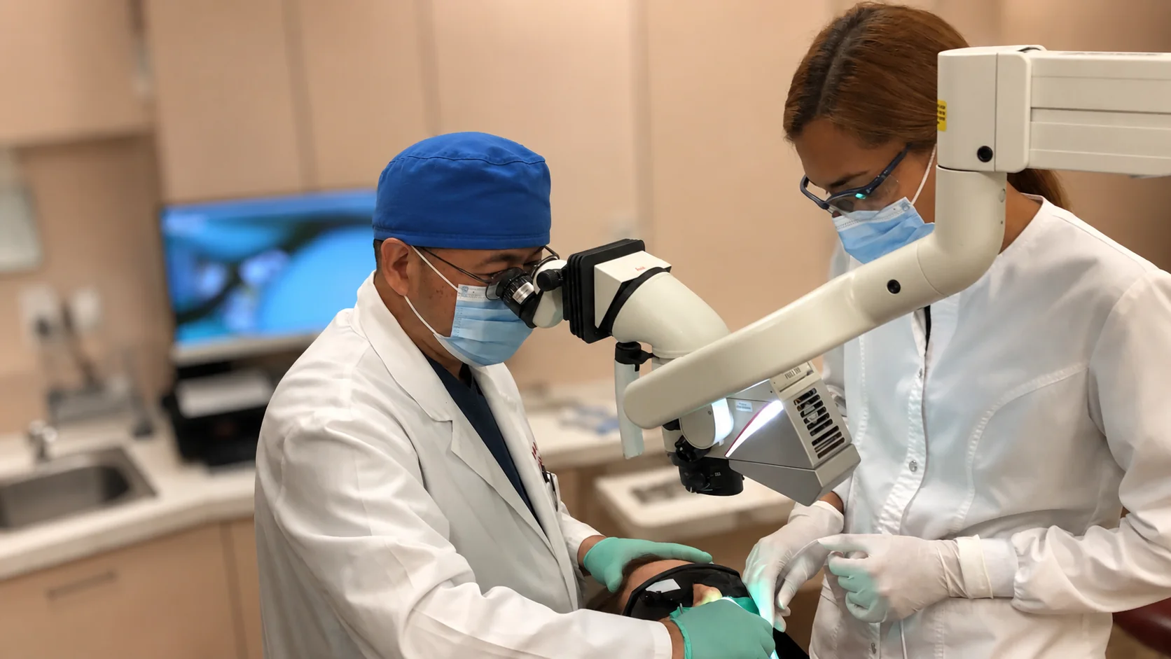
Targeted surgical accessMicroscope-assisted cleaning
Using the Leica microscope, Dr. Nguyen can see the surgical field with magnification and illumination. The infected tissue is removed and the root end is inspected.
Leica microscope precisionRoot-end preparation and filling
The root tip is trimmed, the end of the canal is prepared, and a root-end filling is placed to help seal the canal from the bottom.
Root-end sealHealing and follow-up
Sutures are placed. Over time, the bone can heal around the root end. Follow-up imaging helps monitor healing and confirm that the infection is resolving.
Bone healing over monthsWhy Cone Beam CT Matters for Apicalectomy
A standard dental X-ray is two-dimensional. It can show a problem, but it may not show the exact three-dimensional location, size, or direction of the infection. Cone beam CT provides a 3D view that helps Dr. Nguyen plan with more information before surgery.
Better surgical map
CBCT helps identify the root shape, lesion size, bone thickness, and access path before the procedure begins.
Protect nearby anatomy
For some teeth, CBCT helps show nearby structures such as the sinus, nerve canal, neighboring roots, or thin bone areas.
Better case selection
Not every tooth is a good candidate. CBCT helps Dr. Nguyen decide whether surgery, retreatment, extraction, or another option is safer.
Why a Leica Microscope Helps During Root-End Surgery
Apicalectomy is a small procedure in a very small surgical field. The root end, canal opening, cracks, accessory anatomy, and filling area may be only a few millimeters. Magnification and bright coaxial illumination are important.
Magnified view
The Leica microscope helps Dr. Nguyen see details that are difficult to detect with the naked eye.
Better illumination
Deep or narrow areas need direct light. Microscope lighting helps make the surgical field clearer.
More controlled treatment
Better visualization helps with careful tissue removal, root-end inspection, preparation, and filling placement.
What Patients Usually Feel After Apicalectomy
Many patients expect root-end surgery to be very painful. In reality, post-surgical discomfort is often manageable with proper instructions and medication guidance. Swelling, tenderness, minor bruising, and soreness are possible for several days.
| Timeframe | What may happen | What helps |
|---|---|---|
| First 24 hours | Numbness wears off, tenderness starts, mild bleeding or swelling may occur. | Follow medication instructions, use ice if directed, avoid heavy activity. |
| 2–3 days | Swelling or soreness may peak. The tooth and gum may feel tender. | Eat soft foods, avoid chewing hard foods on that side, keep the area clean as instructed. |
| 1 week | Sutures may be removed or dissolve depending on the type used. | Return for the recommended follow-up appointment. |
| Months later | Bone healing is evaluated over time. Symptoms should improve if healing is progressing. | Keep follow-up visits and X-rays/CBCT if recommended. |
Why Saving the Tooth Can Be Worth It
When a tooth can be predictably saved, keeping the natural tooth may help preserve chewing function, maintain the bite, avoid tooth shifting, and delay or avoid extraction, implant placement, bridge work, or partial denture replacement.
That does not mean apicalectomy is always the right answer. If a tooth has a vertical root fracture, severe periodontal disease, poor remaining structure, or poor long-term prognosis, extraction may be the more honest recommendation. Dr. Nguyen’s goal is not to “do surgery.” The goal is to protect the patient’s long-term dental health.
Questions to Ask Before an Apicalectomy
Is the tooth restorable?
If the tooth cannot be restored with a stable filling or crown, surgery may not be worthwhile.
Is there a crack?
Some cracks make a tooth hopeless. CBCT and microscope evaluation may help, but not every crack is visible before treatment.
Would retreatment be better?
Some cases should be reopened and retreated from inside the canal. Others are better treated surgically from the root end.
What is the backup plan?
If the tooth cannot be saved, ask about extraction, implant, bridge, or partial denture options before deciding.
An apicalectomy is not about doing more dentistry. It is about giving the right tooth one more chance. With cone beam CT, we can study the root and bone in 3D. With the Leica microscope, we can work with better light and magnification. When a tooth is worth saving, precision matters.
— Dr. Minh Nguyen, D.D.S., P.A. · SoftDental HoustonSources and Further Reading
American Association of Endodontists: Endodontic Surgery — explains that during endodontic surgery, a root-end filling may be placed, sutures help the tissue heal, bone heals around the root end over months, and most patients return to normal activities the next day with generally mild discomfort.
American Association of Endodontists: Dental Operating Microscope — explains that microscopes help clinicians view canals, tiny cracks, and problems that might otherwise go unseen.
Leica Microsystems: Dental Surgical Microscopy — describes microscope benefits including light intensity, depth of field, resolution, magnification, and instrument control in deep or narrow cavities.
Still having symptoms after a root canal?
Do not assume the tooth is hopeless.
Dr. Nguyen can evaluate the tooth, review X-rays or cone beam CT findings, and explain whether retreatment, apicalectomy, crown protection, extraction, or another option makes the most sense.
Cone Beam CT · Leica Microscope · Root Canal Therapy · Apical Surgery Evaluation
This article is for patient education only and is not a diagnosis or guarantee of treatment outcome. Apicalectomy candidacy depends on clinical exam, X-rays, CBCT findings, tooth restorability, crack risk, periodontal condition, symptoms, and medical history. Some cases may require referral to an endodontic specialist. © 2026 SoftDental | Dr. Minh Nguyen DDS PA · 10028 West Road Ste. 108, Houston TX 77064 · 281-807-6111
Questions about your own teeth?
Our team is happy to answer them in person, without pressure. Call us or book a visit.
Educational information only. Not a substitute for a personal exam with a licensed dentist.