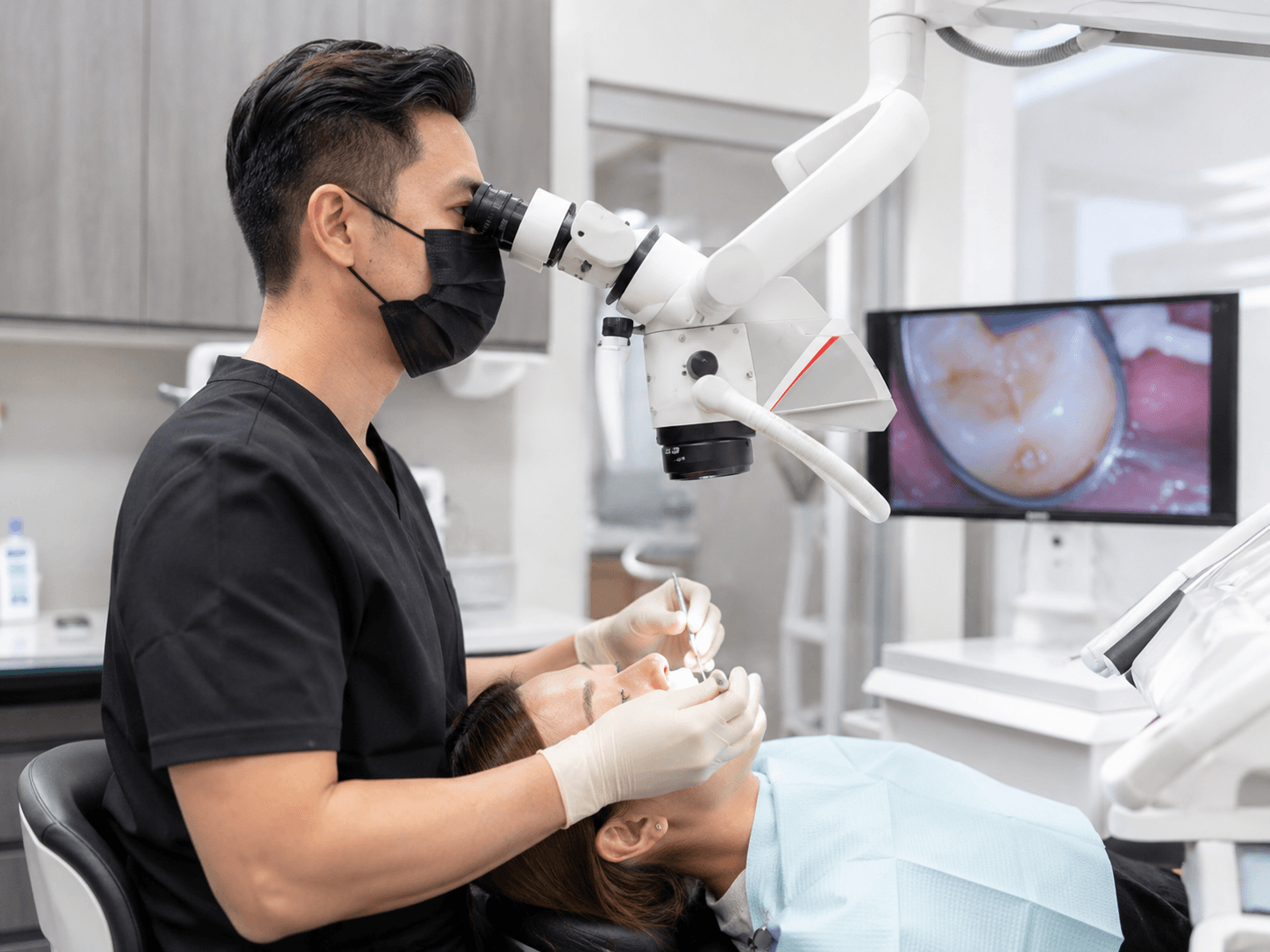
Peri-implantitis remains one of the most challenging complications in implant dentistry. It is a bacterial infection of the tissue and bone around a dental implant — and unlike gum disease around natural teeth, it often progresses faster and with fewer warning signs. The patients who lose implants to peri-implantitis are not careless people. They are people who did not know what was happening until it had gone too far.
What Is Peri-Implantitis?
Peri-implantitis is a pathological condition affecting the tissues around dental implants — characterized by inflammation in the peri-implant tissues and progressive loss of the bone that holds the implant in place. Think of it as gum disease, but specifically around an implant — and more aggressive, because implants lack the natural defense mechanisms that teeth have.
Your natural teeth have a periodontal ligament — a complex tissue structure that acts as a barrier against bacterial invasion. Implants are made of titanium and have no such ligament. Bacteria that accumulate on the implant surface and around the crown can penetrate more directly into the surrounding tissue, which is why peri-implantitis often progresses faster than gum disease around natural teeth.
There are two stages before full peri-implantitis develops:
Healthy Peri-Implant Tissue
The tissue around the implant is healthy — firm, pale pink, no bleeding when probed. This is where every implant should be, and where your daily care and regular check-ups keep it.
- No bleeding when probed or brushed
- Firm, healthy gum around the crown
- Bone level stable on X-ray
- No pain, no bad breath from implant area
Peri-Implant Mucositis — Early Warning (Reversible)
Inflammation is present in the soft tissue only — the bone has not yet been affected. This is the critical window. Peri-implant mucositis may be effectively managed with nonsurgical debridement and control of risk factors. At this stage, the condition is reversible with professional cleaning and improved home care.
- Bleeding on probing or brushing — a key early sign
- Gum redness or puffiness around the crown
- Possible slight bad taste
- No bone loss visible on X-ray yet
Peri-Implantitis — Bone Loss Has Begun (Requires Treatment)
Bacteria have progressed below the gum line and begun destroying the bone around the implant. This is peri-implantitis. The bone loss is visible on X-rays. More advanced cases require individualized surgical approaches. Without treatment, the implant will eventually fail.
- Bleeding, swelling, and possible pus discharge
- Persistent bad breath from implant area
- Crown may appear taller (gum receding)
- Implant may begin to feel loose in advanced cases
- Bone loss visible on X-ray
What Causes Peri-Implantitis?
Peri-implantitis reflects a complex interplay between microbial, host-related, and implant-specific factors. Key systemic and behavioral risk factors include history of periodontitis, smoking, uncontrolled diabetes, poor microbial biofilm control, and obesity, while implant malposition, unfavorable prosthetic factors, and suboptimal peri-implant soft tissue features are relevant site-related factors.
Bacterial Biofilm (Plaque)
The primary driver. Plaque that accumulates on the implant surface and crown margins forms a bacterial colony. Without daily removal, it calcifies into tartar and creates an environment where destructive bacteria thrive — the same pathogens involved in gum disease, but more aggressive around implants.
Smoking
Smoking reduces blood flow to gum tissue, impairs immune response, and dramatically increases peri-implantitis risk. Smokers have significantly higher rates of peri-implantitis and worse outcomes from treatment compared to non-smokers.
History of Gum Disease
Patients who lost teeth to gum disease harbor the same bacterial species in other areas of their mouth. These bacteria can colonize the implant site and initiate peri-implantitis — especially if periodontal disease was not fully treated before implant placement.
Uncontrolled Diabetes
Poorly controlled blood sugar impairs immune function and wound healing, making it harder for the body to fight bacterial infection around the implant. Well-controlled diabetic patients have much better outcomes.
Implant Malposition or Cement Remnants
An implant placed at the wrong angle, too deep, or with excess cement left behind after crown placement creates areas that are impossible to clean — trapping bacteria and providing a permanent source of inflammation.
Skipped Maintenance Visits
Professional cleaning appointments are not optional for implant owners. Even perfect home care cannot remove calcified deposits at the implant collar. These deposits accumulate over skipped appointments and provide the foundation for peri-implantitis to develop.
How Peri-Implantitis Is Treated
Treatment depends entirely on how far the disease has progressed. Early-stage mucositis is reversible with professional care and improved hygiene. Advanced peri-implantitis with significant bone loss requires surgical intervention. The earlier it is caught, the simpler the treatment and the better the outcome.
Non-Surgical Deep Cleaning
For early cases (mucositis or mild peri-implantitis), thorough mechanical debridement — cleaning of the implant surface and surrounding pocket using implant-safe instruments — along with irrigation with antimicrobial agents. Combined with patient home care improvement, this can halt progression and allow healing in early cases.
Antibiotic Therapy
Local or systemic antibiotics may be prescribed alongside mechanical cleaning to reduce the bacterial load. Local delivery inside the implant pocket (a gel or sustained-release chip) targets the specific bacteria at the infection site without the side effects of systemic antibiotics.
Surgical Access and Debridement
For moderate to advanced cases, the gum is opened surgically to provide direct visibility and access to the implant surface. The contaminated surface is meticulously cleaned and decontaminated. Excess bone tissue may be reshaped to eliminate pockets that trap bacteria.
Regenerative Surgery (Bone Grafting)
For cases where significant bone has been lost, regenerative surgery can attempt to rebuild the lost support around the implant using bone graft materials and barrier membranes. Reconstructive surgical approaches appear more consistent, particularly when combined with grafting materials and membranes, based on 2025 clinical reviews.
Photodynamic Therapy (PDT)
An emerging adjunctive treatment: a photosensitive solution is applied to the infected area and activated with a specific wavelength of light, destroying bacteria without damaging tissue. When combined with mechanical debridement, PDT consistently improves probing depth, bleeding on probing, and crestal bone stability.
Supportive Maintenance Program
After treatment, all peri-implantitis cases require a structured maintenance program — typically professional cleanings every 3 months for the first year, then reassessed. Individualized risk-based maintenance protocols substantially enhance clinical outcomes and implant longevity compared with standard fixed-interval maintenance.
I've had patients come to me with bone loss around an implant they did not know was happening — because it had never hurt. That is the lesson of peri-implantitis. The absence of pain does not mean all is well. Regular monitoring is not just a recommendation — for implant patients, it is the difference between a lifelong implant and a failed one.
— Dr. Minh Nguyen, D.D.S., P.A. · SoftDental, Houston TXConcerned about your implant?
If you have noticed bleeding, swelling, or anything unusual around your implant — do not wait. Early intervention is everything. Call SoftDental today.
Educational content only. Statistics cited from peer-reviewed clinical research published through 2025. © 2026 SoftDental | Dr. Minh Nguyen DDS PA · 10028 West Road Ste. 108, Houston TX 77064